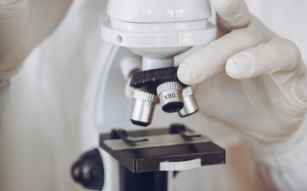
In human history, few instruments have had such a profound impact on our understanding of the world as the microscope. An extraordinary optical device with the ability to magnify objects beyond the limits of human perception.
In this article we will follow the crucial stages of this extraordinary scientific instrument, discovering how the microscope has revolutionized our understanding of the world and opened new frontiers in research.
The invention of the microscope
The first optical microscope was born in 1590 in Holland, in the workshop of Zacharias Jansen and his father Hans, two lens makers who built a rudimentary optical device consisting of convex lenses mounted on a wooden tube.
The real turning point came when, in the 17th century, both Antonie van Leeuwenhoek and Robert Hooke perfected the concept of the microscope. Van Leeuwenhoek, using tiny spherical lenses, was the first to accurately observe and describe bacteria, protozoa, and other microscopic life forms, paving the way for the birth of microbiology. Meanwhile, Hooke, with his microscope composed of multiple lenses, revealed the cellular structure of plants and animal organisms, unveiling a world of complexity hidden from human eyes.
Since then, the microscope has continued to be perfected, eventually achieving lenses capable of correcting more and more optical aberrations, which were impairing observation, allowing higher and more precise magnifications.
The electron microscope revolution
In the 1930s the electron microscope saw the light of day, which, unlike the optical device that uses a beam of light, uses a beam of electrons that strikes the object to be observed and then reconstructs its image.
Because of limitations in resolving power resulting from the physical nature of glass lenses, efforts have been made to overcome this obstacle by adopting magnetic lenses. These lenses allow shorter wavelengths to pass through, thus improving the resolution of details. This is particularly useful for exploring the intrinsic nature of observed objects and revealing the presence of pathogens such as viruses, which in the past could only be assumed and still elude observation with the traditional light microscope.
The electron microscope has continued to evolve over the years, with one technique in particular proving so revolutionary that it earned the Nobel Prize in Chemistry in 2017. We are talking about cryogenic transmission electron microscopy (Cryo-TEM), which combines sophisticated sample preparation with the use of a powerful electron microscope and advanced image analysis software.
The discovery by Jacques Dubochet, Joachim Frank and Richard Henderson made it possible to visualize each individual structure with a resolution of a few billionths of a meter, enabling scientists to explore matter atom by atom and obtain the three-dimensional structure of biological macromolecules such as DNA, RNA and proteins.
The cryoelectron microscopy technique has also made it possible to observe fragile structures that would otherwise have been damaged by electron impact. In 2017, for example, Cryo-TEM was successfully used to reconstruct the complete three-dimensional structure of the Zika virus.
What is the future of microscopy?
Research in the field of microscopy is varied, with many aspects amenable to improvement beyond simply enhancing resolving power. Among the most notable challenges:
- New types of dyes and calibration slides, enabling improved image definition and acquisition.
- Improved techniques to avoid damage to sensitive samples while maintaining the ability to interact with them.
- Introduction of automations and artificial intelligence to improve the capabilities of microscopes.
Artificial intelligence, in particular, is the latest frontier in the world of microscopy, with manufacturers continuing to implement and refine AI and automation systems in their microscopes. This allows them to obtain faster images, reduce sample preparation time and introduce new features.
The world of microscopy is vast and complex, and in this article we have provided only a general overview of one of the most significant instruments in the history of scientific research.